
Contents
Dental Granuloma: Causes, Symptoms, Treatment Options, and Costs in 2026
A dental granuloma is one of those conditions that can silently threaten your oral health for months or even years before you realize something is wrong. Because granulomas are frequently painless in their early stages, they are often discovered incidentally on routine dental X-rays -- underscoring the importance of regular dental check-ups. According to the American Association of Endodontists (AAE), periapical lesions including granulomas account for approximately 75% of all inflammatory lesions affecting the jawbone.
In this comprehensive guide, we explain what a dental granuloma is, how it forms, the symptoms to watch for, all available treatment options, and what American patients can expect to pay for treatment in 2026. Whether you have been newly diagnosed or are trying to understand a loved one's condition, this article provides the clinical knowledge you need to make informed decisions.
What Is a Dental Granuloma?
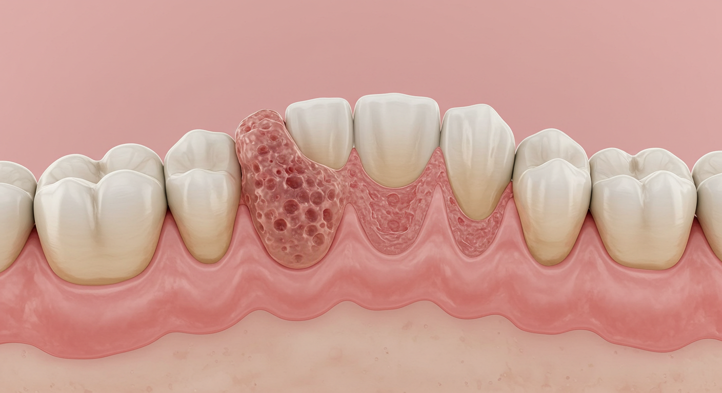
A dental granuloma -- medically known as a periapical granuloma or apical granuloma -- is a small, rounded mass of chronically inflamed tissue that forms at the tip (apex) of a tooth root. It develops as the body's immune response to a persistent bacterial infection originating from the tooth's interior (the pulp chamber and root canals).
When bacteria invade the dental pulp -- through a deep cavity, crack, or trauma -- the pulp tissue dies (necrosis). The bacteria then migrate through the root canal system to the apex, where they exit into the surrounding bone tissue. Rather than allowing the infection to spread unchecked, the body walls off the bacteria by forming a granuloma -- a sphere of immune cells (macrophages, lymphocytes, and plasma cells) surrounded by fibrous tissue.
A typical dental granuloma ranges from 2mm to 10mm in diameter. Lesions larger than 10mm are more likely to be classified as periapical cysts, which have a fluid-filled center and may require different treatment approaches.
"A periapical granuloma is essentially the body's attempt to contain a chronic infection. Think of it as a biological quarantine zone -- the immune system builds a wall of inflammatory tissue to prevent bacteria from spreading into the jawbone and bloodstream. While this containment strategy works temporarily, it is not a cure, and without treatment, the infection will eventually overwhelm the body's defenses."
Symptoms and Warning Signs
One of the most challenging aspects of dental granulomas is that they are often completely asymptomatic in the early and even intermediate stages. Many patients are surprised to learn they have a granuloma because they feel no pain. However, certain subtle signs can indicate the presence of a granuloma:
- Mild, intermittent discomfort -- A vague ache or pressure sensation near the affected tooth, especially when biting down or applying pressure. This discomfort may come and go over weeks or months.
- Tooth discoloration -- A darkening, graying, or yellowing of the affected tooth, which indicates that the pulp has undergone necrosis (death). This is one of the earliest visible signs.
- Localized gum swelling -- A small, persistent bump or fullness in the gum tissue near the root tip. This may feel firm to the touch and is usually not painful initially.
- Sensitivity to hot or cold -- Early-stage pulp involvement may produce lingering sensitivity, though once the pulp dies completely, hot/cold sensitivity typically disappears.
- Bad taste or odor -- If the granuloma develops a draining sinus tract (fistula), you may notice a persistent bad taste in your mouth or a small pimple-like bump on the gum that oozes pus intermittently.
- Tooth mobility -- In advanced cases where significant bone loss has occurred, the affected tooth may become slightly loose.
Warning: The absence of pain does not mean the condition is harmless. Many dental granulomas grow silently for years, causing progressive bone destruction. By the time pain develops, the infection has often advanced to an abscess stage requiring more aggressive (and expensive) treatment. This is why the ADA recommends dental X-rays at regular intervals even when you have no symptoms.
How Is a Dental Granuloma Diagnosed?
Diagnosing a dental granuloma requires professional dental evaluation. Self-diagnosis is virtually impossible because the lesion exists beneath the gum tissue and inside the bone. Here is how your dentist or endodontist will identify a granuloma:
- Periapical X-ray (radiograph) -- The primary diagnostic tool. A granuloma appears as a well-defined, dark (radiolucent) area at the tip of the tooth root, typically round or oval in shape. This dark zone represents the loss of normal bone density where the inflamed tissue resides.
- Cone-beam CT scan (CBCT) -- For complex cases or when the standard X-ray is inconclusive, a 3D CBCT scan provides detailed cross-sectional images that reveal the exact size, shape, and extent of the lesion, as well as its relationship to adjacent anatomical structures.
- Pulp vitality testing -- The dentist uses cold, electric, or thermal stimuli to test whether the tooth's nerve is alive or dead. A tooth with a granuloma typically shows no response (necrotic pulp).
- Percussion and palpation -- Tapping on the tooth (percussion) or pressing on the gum tissue over the root tip (palpation) may produce tenderness or discomfort.
- Histopathological examination -- The definitive distinction between a granuloma and a periapical cyst can only be made through microscopic examination of the tissue after it has been removed. However, this is usually not necessary for treatment planning, since both conditions are treated similarly.
Good to Know: The ADA recommends that adults with existing dental restorations receive periapical or bitewing X-rays every 12 to 24 months. For adults at higher risk of dental disease (smokers, diabetics, patients with a history of decay), annual radiographs are recommended. These routine films are your first line of defense against undetected granulomas.
Root Causes of Dental Granulomas
Dental granulomas are always the result of bacterial infection reaching the periapical tissues through the root canal system. The four most common pathways for this infection are:
- Untreated dental cavities -- A deep cavity that penetrates through the enamel and dentin layers eventually reaches the pulp chamber, introducing bacteria that cause pulp necrosis and subsequent granuloma formation. This is the most common cause.
- Failed or incomplete root canal treatment -- If a previous root canal procedure did not fully eliminate bacteria from all canals (missed canals, inadequate cleaning, or poor obturation), the residual infection can lead to a granuloma. Studies suggest that 5-15% of primary root canal treatments fail over a 10-year period.
- Dental trauma -- A blow to a tooth from a sports injury, fall, or accident can sever the blood supply to the pulp, causing it to die. The resulting necrotic tissue becomes a breeding ground for bacteria that enter through microcracks in the root.
- Advanced periodontal disease -- In severe periodontitis, bacteria can reach the tooth root apex through deep periodontal pockets, creating a combined endodontic-periodontic lesion that requires complex treatment.
"In my practice, I see dental granulomas most commonly in patients who delayed treatment for a symptomatic cavity. What started as a small, easily treatable cavity eventually became a root canal problem. By the time they come to me, the granuloma may have been growing silently for a year or more. The lesson is clear: treat cavities early before they become endodontic emergencies."
Treatment Options for Dental Granuloma
The treatment approach for a dental granuloma depends on the tooth's condition, the size of the lesion, and whether prior endodontic treatment has been attempted. Here are the main options available in the US:
1. Root Canal Therapy (Primary Endodontic Treatment) -- This is the first-line treatment for most granulomas. The endodontist or general dentist removes the infected pulp tissue, thoroughly cleans and disinfects all root canals using specialized instruments and irrigating solutions (typically sodium hypochlorite), shapes the canals, and fills them with a biocompatible material called gutta-percha. The tooth is then sealed with a permanent crown or restoration. Success rate: approximately 85-95% for primary root canals.
2. Endodontic Retreatment -- If a previously root-canal-treated tooth develops a granuloma (indicating treatment failure), the existing filling material is removed, the canals are re-cleaned and re-shaped, and new filling material is placed. This is more complex than primary treatment and has a slightly lower success rate of 75-85%.
3. Apicoectomy (Surgical Endodontics) -- When conventional root canal therapy fails or is not feasible, an apicoectomy is performed. The endodontist makes a small incision in the gum, accesses the root tip through the bone, removes the infected root tip (apex) along with the surrounding granulomatous tissue, and places a retrograde filling to seal the root end. Success rate: approximately 90-95%.
4. Tooth Extraction -- When the tooth is severely compromised (vertical root fracture, extensive decay, insufficient remaining tooth structure), extraction may be the only option. Following extraction, the patient can pursue replacement options including a dental implant, bridge, or removable prosthesis.
5. Antibiotic Therapy (Adjunctive) -- Antibiotics alone cannot cure a dental granuloma because the infection source (necrotic pulp) remains. However, antibiotics such as amoxicillin or clindamycin may be prescribed before or after the procedure to control acute flare-ups, reduce swelling, or prevent systemic spread in immunocompromised patients.
Cost of Dental Granuloma Treatment in the US
Treatment costs vary significantly based on the tooth location, complexity, provider, and geographic region. Here are typical costs American patients can expect in 2026:
| Treatment | Cost Without Insurance | Typical Insurance Coverage | Est. Out-of-Pocket |
|---|---|---|---|
| Root canal (anterior tooth) | $700 - $1,100 | 50-80% after deductible | $140 - $550 |
| Root canal (premolar) | $800 - $1,300 | 50-80% after deductible | $160 - $650 |
| Root canal (molar) | $1,000 - $1,800 | 50-80% after deductible | $200 - $900 |
| Endodontic retreatment | $1,000 - $2,000 | 50-80% after deductible | $200 - $1,000 |
| Apicoectomy | $900 - $1,500 | 50-80% after deductible | $180 - $750 |
| Dental crown (after root canal) | $800 - $1,800 | 50% after deductible | $400 - $900 |
| Extraction (surgical) | $250 - $600 | 50-80% after deductible | $50 - $300 |
| CBCT scan | $150 - $450 | Varies widely | $75 - $450 |
Good to Know: Most dental insurance plans classify root canal therapy as a "major" procedure covered at 50-80% after deductible, subject to the plan's annual maximum (typically $1,000-$2,500). If you lack insurance, many endodontists offer payment plans through CareCredit or Lending Club, and dental schools affiliated with universities often provide endodontic treatment at 40-60% below private practice rates.
Complications of Untreated Dental Granulomas
While a dental granuloma may seem harmless because it is often painless, leaving it untreated can lead to serious complications that affect both oral and general health:
- Periapical cyst formation -- A granuloma can evolve into a periapical cyst, a fluid-filled sac lined with epithelium that can grow significantly larger, causing more extensive bone destruction and potentially affecting adjacent teeth.
- Dental abscess -- The chronic infection can flare into an acute abscess with severe pain, facial swelling, fever, and difficulty opening the mouth. Dental abscesses require emergency treatment.
- Sinus tract (fistula) -- The body may create a drainage pathway from the granuloma through the bone and gum tissue, resulting in a persistent pimple-like bump on the gum that periodically drains pus.
- Jawbone destruction -- Progressive bone loss around the root tip weakens the jaw and can compromise the stability of neighboring teeth.
- Systemic spread of infection -- In rare but serious cases, bacteria from a dental granuloma can enter the bloodstream (bacteremia), potentially affecting the heart valves (endocarditis), brain, or other organs. Patients with compromised immune systems, heart valve disorders, or artificial joints are at elevated risk.
Warning: If you experience sudden, severe facial swelling accompanied by fever, difficulty swallowing or breathing, or swelling that extends to the eye or neck area, seek emergency medical attention immediately. These are signs that a dental infection has spread beyond the local area and can become life-threatening if not treated promptly. Go to an emergency room -- do not wait for a dental appointment.
Prevention Strategies for Dental Granulomas
Preventing dental granulomas centers on preventing the conditions that allow bacteria to reach the root apex. Here are evidence-based strategies:
- Maintain thorough oral hygiene -- Brush twice daily with ADA-accepted fluoride toothpaste, floss or use a water flosser daily, and consider an antimicrobial mouthwash. Good hygiene prevents cavities, which are the leading cause of pulp infection.
- Treat cavities promptly -- A small cavity treated with a filling costs $150-$300. A cavity that reaches the pulp requires a root canal costing $700-$1,800 plus a crown. Early treatment is both healthier and significantly cheaper.
- Schedule regular dental check-ups -- The ADA recommends at least one dental visit per year, with periodic X-rays to detect problems invisible to the naked eye. Many dentists recommend twice-yearly visits.
- Wear a mouthguard during sports -- Dental trauma is a leading cause of pulp necrosis in young adults. Custom-fitted mouthguards from your dentist provide the best protection.
- Address bruxism (teeth grinding) -- Chronic grinding can crack teeth, creating pathways for bacterial invasion. A night guard can prevent this damage.
- Limit sugar intake -- The ADA reports that frequent sugar consumption is the primary dietary risk factor for dental decay. Reducing sugary snacks and beverages lowers your cavity risk substantially.
| Prevention Action | Estimated Annual Cost | Potential Savings vs. Treatment |
|---|---|---|
| Fluoride toothpaste + floss | $30 - $60 | Up to $3,000+ (root canal + crown) |
| Two dental check-ups + cleanings | $200 - $500 | Early detection prevents complex procedures |
| Filling a small cavity | $150 - $300 | $1,500 - $3,000+ in avoided root canal costs |
| Custom sports mouthguard | $300 - $500 (one-time) | Prevents trauma-related root canals |
Frequently Asked Questions About Dental Granulomas
Is a dental granuloma dangerous if left untreated?
Yes. While not immediately life-threatening in most cases, an untreated dental granuloma will continue to grow, destroying surrounding bone tissue. It can evolve into a periapical cyst, flare into an acute dental abscess with severe pain and facial swelling, or in rare but documented cases, allow bacteria to enter the bloodstream, posing risks of endocarditis, brain abscess, or sepsis. The AAE strongly recommends treating all diagnosed granulomas promptly.
Can a dental granuloma heal on its own without treatment?
No. A dental granuloma cannot resolve on its own because the source of the infection -- the necrotic pulp tissue inside the tooth -- remains. The body's immune response can contain the infection temporarily (which is what the granuloma represents), but it cannot eliminate the bacteria within the sealed root canal system. Only professional endodontic treatment or extraction can remove the infection source and allow the bone to heal.
How long does it take to recover from granuloma treatment?
Recovery from a standard root canal procedure is relatively quick -- most patients return to normal activities within 24-48 hours. Mild soreness at the treatment site is common for 3-5 days and can be managed with over-the-counter pain relievers like ibuprofen. Recovery from an apicoectomy takes slightly longer, typically 7-14 days for soft tissue healing, though complete bone regeneration around the root tip may take 6-12 months, which is monitored through follow-up X-rays.
Does dental insurance cover granuloma treatment?
Most dental insurance plans cover root canal therapy as a "major" procedure at 50-80% of the allowed amount, after the annual deductible has been met. However, coverage is subject to the plan's annual maximum benefit, which is typically $1,000-$2,500. If your granuloma requires both a root canal and a crown, the combined cost may exceed your annual maximum. FSA and HSA funds can be used to cover out-of-pocket dental expenses, and many endodontists accept CareCredit financing.
Can a dental granuloma come back after treatment?
While recurrence is possible, it is relatively uncommon after properly performed endodontic treatment. Primary root canal therapy has a success rate of 85-95%, and apicoectomy succeeds in approximately 90-95% of cases. Recurrence typically indicates that bacteria were not fully eliminated during the initial treatment -- for example, a missed canal or an accessory canal that was not cleaned. If a granuloma recurs, retreatment or apicoectomy can address the issue. Regular follow-up X-rays at 6 months and 1 year post-treatment are standard to confirm healing.
"The prognosis for dental granulomas treated with modern endodontic techniques is excellent. With today's rotary instrumentation, advanced irrigation protocols, and cone-beam CT imaging, we can achieve predictable outcomes that save the natural tooth in the vast majority of cases. Saving a tooth is almost always preferable to extraction -- your natural tooth, when properly restored, will serve you better than any implant or bridge."
Sources
- 1. American Association of Endodontists. "Periapical Lesions: Diagnosis and Treatment Guidelines." AAE Clinical Practice Guidelines, 2024.
- 2. Nair PNR. "Pathogenesis of Apical Periodontitis and the Causes of Endodontic Failures." Critical Reviews in Oral Biology & Medicine, 2004;15(6):348-381.
- 3. American Dental Association. "Dental Radiographic Examinations: Recommendations for Patient Selection and Limiting Radiation Exposure." ADA.org, 2024.
- 4. Torabinejad M, et al. "Outcomes of Nonsurgical Retreatment and Endodontic Surgery: A Systematic Review." Journal of Endodontics, 2009;35(7):930-937.
- 5. Fair Health Consumer. "Root Canal and Endodontic Treatment Cost Estimates." FairHealthConsumer.org, 2025.
- 6. National Association of Dental Plans. "Dental Benefits Coverage: Annual Report." NADP Research Brief, 2025.
- 7. Von Arx T, et al. "Success Rates of Apicoectomy: A Meta-Analysis." International Endodontic Journal, 2020;53(2):166-185.
- 8. Centers for Disease Control and Prevention. "Oral Health Surveillance: Dental Caries and Tooth Loss in Adults." CDC.gov, 2024.