
Contents
Necrotic Tooth: Symptoms, Causes, and Treatment Options
A necrotic tooth -- commonly called a "dead tooth" -- is a tooth whose internal pulp tissue has died due to infection, trauma, or loss of blood supply. According to the American Association of Endodontists (AAE), over 15 million root canal procedures are performed in the United States each year, and dental necrosis is one of the leading reasons patients need endodontic treatment. Left untreated, a necrotic tooth can lead to serious infections including dental abscesses, bone loss, and in rare cases, life-threatening systemic complications.
This guide provides a thorough explanation of what dental necrosis is, how to recognize its symptoms, what causes it, what treatment options are available, and how much treatment costs in the US.
What Is a Necrotic Tooth?
Every tooth contains a soft tissue center called the dental pulp. The pulp is located inside the pulp chamber (in the crown) and the root canals (in the roots). It contains blood vessels, nerves, and connective tissue that nourish the tooth and provide sensation. When this living tissue dies -- whether from bacterial infection, physical trauma, or interrupted blood flow -- the tooth is said to be necrotic.
A necrotic tooth is no longer vital (alive), but it can remain structurally intact in the jaw for years. However, without treatment, the dead pulp tissue becomes a breeding ground for bacteria, which can spread beyond the tooth into the surrounding bone and soft tissues.
"Many patients are surprised to learn that a tooth can die silently. The initial pain from pulpitis may subside, leading patients to believe the problem resolved itself. In reality, the nerve has died, and the infection is spreading without symptoms. This is why regular dental checkups are so critical."
How Dental Pulp Dies: The Stages of Necrosis
Pulp necrosis typically progresses through several stages:
- Reversible pulpitis: The pulp becomes inflamed due to a shallow cavity or minor trauma. Pain occurs with stimuli (hot, cold, sweet) but resolves quickly. At this stage, treatment (a filling or crown) can save the pulp.
- Irreversible pulpitis: Inflammation becomes severe and cannot resolve on its own. The patient experiences spontaneous, intense, lingering pain -- the classic "raging toothache." The pulp is dying.
- Partial necrosis: Some pulp tissue has died, but portions may still be alive. Pain may become intermittent or change character.
- Complete necrosis: The entire pulp is dead. Pain may temporarily disappear (because the nerve is destroyed), creating a dangerous false sense of relief. The tooth may darken in color.
- Periapical infection: Bacteria from the necrotic pulp leak through the root tip into the surrounding bone, forming a periapical abscess or granuloma. Pain, swelling, and sometimes fever return.
Warning: A sudden disappearance of severe tooth pain does NOT mean the problem is resolved. It often means the nerve has died. The infection continues to spread silently. See your dentist immediately if you experience this pattern.
Symptoms of a Necrotic Tooth
The symptoms of a necrotic tooth can be confusing because they change as the condition progresses. Common signs include:
- Tooth discoloration: The tooth turns gray, dark yellow, or black as the internal blood supply degrades and breakdown products stain the dentin from within.
- History of severe pain that suddenly stopped: This pattern suggests the nerve has died.
- Sensitivity or no sensitivity to temperature: A necrotic tooth typically does not respond to hot or cold stimuli, though an early-stage tooth may still react.
- Swelling of the gum near the tooth: A small bump (fistula or "gumboil") may appear on the gum, indicating a draining abscess.
- Bad taste or odor: Pus draining from an abscess through a fistula can cause a foul taste in the mouth.
- Pain when biting or pressing on the tooth: Periapical inflammation makes the tooth tender to pressure.
- Facial swelling: In severe cases, infection can spread to the surrounding tissues, causing visible swelling of the cheek, jaw, or under the eye.
Good to Know: A necrotic tooth does not always cause pain. Many dead teeth are discovered incidentally during routine dental X-rays, which may show a dark shadow (radiolucency) around the root tip indicating chronic infection. This is why the ADA recommends dental checkups every 6 months.
What Causes Dental Necrosis?
Four primary pathways can lead to pulp necrosis:
| Cause | Mechanism | Prevalence |
|---|---|---|
| Deep dental cavity | Bacteria penetrate through enamel and dentin to infect the pulp, causing pulpitis progressing to necrosis | Most common (~60% of cases) |
| Dental trauma | A blow, fall, or sports injury severs the blood vessels at the root tip, cutting off blood supply to the pulp | ~25% of cases |
| Periodontal disease | Bacteria from deep periodontal pockets enter the pulp through lateral canals or the root apex | ~10% of cases |
| Iatrogenic causes | Dental procedures (deep fillings, crown prep, aggressive whitening) that generate excessive heat or remove too much tooth structure near the pulp | ~5% of cases |
"Trauma-related necrosis is particularly insidious because it can occur months or even years after the initial injury. I have seen patients who experienced a sports impact in high school present with a necrotic tooth in their thirties. Any tooth that has suffered trauma should be monitored with periodic vitality testing and X-rays."
Diagnosing a Necrotic Tooth
Your dentist or endodontist uses a combination of tests to confirm pulp necrosis:
- Thermal testing: A cold stimulus (often a frozen cotton pellet with tetrafluoroethane refrigerant spray) is applied to the tooth. A vital tooth responds with a sharp sensation; a necrotic tooth has no response.
- Electric pulp testing (EPT): A small electrical current is applied to the tooth surface. A vital pulp will respond at a certain threshold; a necrotic tooth shows no response at maximum stimulation.
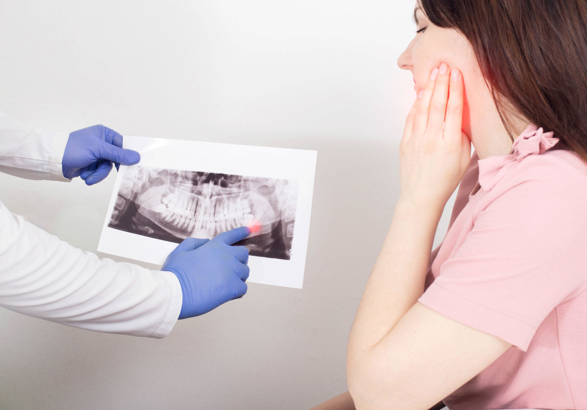
- Periapical X-ray: Reveals a dark area (radiolucency) around the root tip, indicating bone loss from chronic infection.
- Percussion test: Tapping on the tooth may produce pain if periapical inflammation is present.
- CBCT (cone-beam CT) scan: In complex cases, 3D imaging provides detailed views of the root anatomy and the extent of periapical pathology.
- Visual examination: Discoloration, swelling, or a draining fistula are visible indicators.
Treatment Options for a Necrotic Tooth
Once a tooth is confirmed necrotic, the dead tissue must be removed to eliminate the source of infection. There are three main treatment pathways:
Root Canal Therapy: The Primary Treatment
Root canal therapy (endodontic treatment) is the standard of care for saving a necrotic tooth. The procedure involves:
- Anesthesia: Local anesthetic numbs the tooth and surrounding area. Even though the pulp is dead, surrounding tissues can still feel pain.
- Access opening: The dentist drills through the crown of the tooth to reach the pulp chamber.
- Pulp removal: Specialized endodontic files are used to remove all necrotic tissue from the pulp chamber and root canals.
- Cleaning and shaping: The canals are cleaned with antimicrobial irrigants (typically sodium hypochlorite) and shaped to a specific taper for optimal filling.
- Obturation: The cleaned canals are filled with gutta-percha (a biocompatible rubber-like material) and sealer cement to create a hermetic seal.
- Restoration: A permanent filling is placed, and in most cases, a dental crown is recommended to protect the weakened tooth from fracture.
Root canal success rates are excellent -- the AAE reports a success rate of approximately 95% for initial root canal treatments performed by endodontists.
Apicoectomy: When Root Canal Alone Is Not Enough
If a root canal fails to resolve the infection -- or if retreatment through the crown is not feasible due to a post, complex anatomy, or persistent pathology -- an apicoectomy (root-end surgery) may be performed. This microsurgical procedure involves accessing the root tip through the gum and bone, removing the infected tip and surrounding tissue, and placing a retrograde filling to seal the root end.
Extraction as a Last Resort
When a tooth is too damaged to restore -- for example, if the root is fractured, the tooth has extensive decay below the bone level, or multiple previous treatments have failed -- extraction may be the only remaining option. After extraction, the missing tooth can be replaced with a dental implant, bridge, or removable prosthesis.
Good to Know: Saving a natural tooth with a root canal is almost always preferable to extraction. A root canal preserves the natural tooth structure, maintains bone density, prevents adjacent teeth from shifting, and costs less than extraction followed by implant placement (which can total $4,000 - $6,000+).
Cost of Treating a Necrotic Tooth in the US
The cost of treating a necrotic tooth depends on the tooth's location, the complexity of the root canal system, and the type of restoration needed afterward. Here are typical US costs:
| Procedure | Front Tooth (Incisor/Canine) | Premolar | Molar |
|---|---|---|---|
| Root canal (general dentist) | $700 - $1,100 | $800 - $1,200 | $1,000 - $1,600 |
| Root canal (endodontist) | $900 - $1,500 | $1,000 - $1,800 | $1,200 - $2,200 |
| Dental crown (porcelain) | $1,000 - $2,000 | $1,000 - $2,000 | $1,000 - $2,500 |
| Apicoectomy | $900 - $1,500 | $1,000 - $1,700 | $1,200 - $2,000 |
| Extraction (surgical) | $200 - $600 | $250 - $700 | $300 - $900 |
| Total (root canal + crown) | $1,700 - $3,100 | $1,800 - $3,200 | $2,000 - $4,700 |
Insurance Coverage and Financing
Most dental insurance plans classify root canals as a "major" or "basic" service, typically covered at 50-80% after the deductible, up to the plan's annual maximum (usually $1,000 - $2,500 per year). Crowns are typically classified as "major" restorative and covered at 50%. Here are strategies to manage the out-of-pocket cost:
- Verify your insurance coverage: Call your insurer before treatment to confirm your benefit level for root canals and crowns, and check how much of your annual maximum remains.
- Use HSA / FSA funds: Pre-tax health savings account or flexible spending account dollars can cover endodontic treatment, reducing your effective cost by 20-35%.
- Payment plans: Many dental offices offer interest-free monthly payment plans. Third-party financing through CareCredit or LendingClub is also widely available.
- Dental schools: University dental clinics offer root canals performed by endodontic residents under faculty supervision at 30-50% lower cost than private practice.
Complications of Untreated Dental Necrosis
Ignoring a necrotic tooth is dangerous. Without treatment, the infection can progress to increasingly serious complications:
- Periapical abscess: A pus-filled pocket forms at the root tip, causing severe pain, swelling, and fever.
- Granuloma or cyst: A chronic inflammatory mass develops around the root tip, slowly destroying surrounding bone.
- Cellulitis: The infection spreads to the soft tissues of the face and neck, causing dangerous swelling that can compromise the airway.
- Ludwig's angina: A life-threatening infection of the floor of the mouth that can obstruct breathing. This is a medical emergency.
- Osteomyelitis: The infection spreads to the jawbone itself, requiring prolonged antibiotic therapy and sometimes surgery.
- Sepsis: In rare cases, oral bacteria can enter the bloodstream, causing sepsis -- a systemic infection that can be fatal without emergency treatment.
- Cavernous sinus thrombosis: In extremely rare cases involving upper teeth, infection can spread to the brain's cavernous sinus, which is life-threatening.
Warning: If you experience facial swelling, difficulty swallowing, difficulty breathing, high fever, or swelling that is rapidly spreading from a dental infection, go to the emergency room immediately. These are signs of a potentially life-threatening infection that requires emergency medical intervention, not just dental treatment.
Prevention: How to Protect Your Teeth From Necrosis
While not all cases of dental necrosis can be prevented (especially trauma-related), these evidence-based strategies significantly reduce your risk:
- Maintain excellent oral hygiene: Brush twice daily with fluoride toothpaste and floss daily to prevent cavities from reaching the pulp.
- Visit your dentist every 6 months: Regular checkups and professional cleanings catch cavities early, before they penetrate deep enough to threaten the pulp.
- Treat cavities promptly: A small filling today prevents a root canal tomorrow. Never ignore a dentist's recommendation to treat a cavity.
- Wear a mouthguard during sports: Custom-fitted athletic mouthguards (available from your dentist for $200-$500) dramatically reduce the risk of dental trauma during contact sports.
- Address bruxism: Chronic teeth grinding can cause microfractures that allow bacteria to reach the pulp. A night guard can protect your teeth.
- Limit sugary and acidic foods: Sugar feeds cavity-causing bacteria; acid erodes protective enamel. Both increase your risk of pulp exposure.
- Monitor previously traumatized teeth: If a tooth has ever been hit or injured, inform your dentist so it can be monitored with periodic vitality tests.
FAQ About Necrotic Teeth
Does a necrotic tooth always hurt?
No. Pain is not a reliable indicator of dental necrosis. In the early stages (pulpitis), there is often severe pain. But once the nerve dies completely, the pain may temporarily disappear for weeks, months, or even years. During this "quiet" period, the infection continues spreading silently in the bone. Pain typically returns when a periapical abscess forms. Some necrotic teeth are discovered only through routine X-rays with no pain at all.
What happens if you ignore a necrotic tooth?
The infection will not resolve on its own. Over time, bacteria from the dead pulp will spread through the root tip into the surrounding jawbone, forming an abscess, granuloma, or cyst. This causes bone destruction around the root. Eventually, the infection can spread to the soft tissues of the face and neck (cellulitis), potentially compromising the airway. In rare but documented cases, untreated dental infections have led to sepsis and death. An untreated necrotic tooth will ultimately be lost.
Can a dead tooth be saved?
Yes, in most cases. Root canal therapy successfully saves necrotic teeth approximately 95% of the time when performed by an endodontist. The dead pulp is removed, the canals are cleaned and sealed, and the tooth is restored with a crown. The treated tooth can function normally for decades -- often for the rest of the patient's life. The tooth is no longer "alive" (it has no nerve or blood supply), but it remains structurally sound and fully functional.
How long can a necrotic tooth stay in your mouth?
A necrotic tooth can remain in the mouth for months or years without causing obvious symptoms, but the infection is progressing the entire time. Bone is being destroyed around the root, and the risk of acute flare-ups (abscesses) and systemic complications increases with time. There is no safe period for leaving a necrotic tooth untreated. The sooner you receive root canal therapy, the better the outcome and the less bone loss you will experience.
Is a root canal painful?
Modern root canal therapy is performed under local anesthesia and is generally no more uncomfortable than getting a filling. A survey by the AAE found that patients who have actually had a root canal are six times more likely to describe it as "painless" compared to patients who have never had one. After the procedure, mild soreness for 2-3 days is normal and can be managed with over-the-counter pain relievers like ibuprofen. The procedure relieves the pain of the infection -- it does not cause pain.
Sources
- 1. American Association of Endodontists. "Root Canal Treatment." AAE.org, 2025.
- 2. Siqueira JF, Rocas IN. "Clinical implications and microbiology of bacterial persistence after treatment procedures." J Endod. 2008;34(11):1291-1301.
- 3. Ng YL, Mann V, Gulabivala K. "Tooth survival following non-surgical root canal treatment: a systematic review of the literature." Int Endod J. 2010;43(3):171-189.
- 4. American Dental Association. "Root Canals." ADA.org, 2025.
- 5. Berman LH, Hargreaves KM. "Cohen's Pathways of the Pulp." 12th ed. Elsevier, 2021.
- 6. Fouad AF. "Endodontic Microbiology." 2nd ed. Wiley, 2017.
- 7. AAE Patient Survey. "Perceptions of Root Canal Treatment." AAE, 2024.
- 8. Segura-Egea JJ, et al. "Endodontic medicine: connections between apical periodontitis and systemic diseases." Int Endod J. 2015;48(10):933-951.
- 9. Consumer Guide to Dentistry. "Root Canal Cost." yourdentistryguide.com, 2025.