
Contents
Cavity vs Stain on Teeth: How to Tell the Difference in 2026
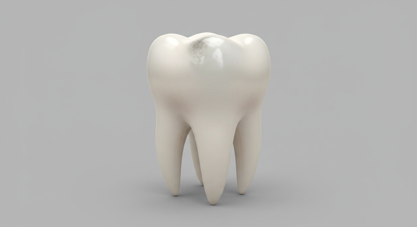
You glance in the mirror and notice a dark spot on one of your teeth. Your first instinct is to wonder whether it is a cavity that needs urgent attention or a harmless stain you can manage at home. This is one of the most common dental concerns among Americans -- the ADA estimates that roughly 90 percent of adults have had at least one cavity by age 65, while surface stains affect virtually everyone who drinks coffee, tea, or red wine. Knowing the difference between a cavity and a stain can save you time, money, and potentially your tooth.
In this comprehensive 2026 guide, we break down the visual, sensory, and clinical differences between cavities and stains, review the latest diagnostic tools now available in dental offices across the United States, compare treatment costs and insurance coverage, and outline proven prevention strategies that actually work.
Understanding Cavities and Stains
What Exactly Is a Dental Cavity?
A dental cavity, also called dental caries or tooth decay, is a permanent structural breakdown of the tooth's enamel, dentin, or cementum. It begins when bacteria in dental plaque metabolize sugars and starches from food, producing acids that demineralize the hard outer layer of the tooth. Over time, this acid attack creates a hole -- the cavity -- that progressively deepens if left untreated.
According to the Centers for Disease Control and Prevention (CDC), untreated cavities affect about 26 percent of U.S. adults between 20 and 64 years of age. Cavities can form on any surface of a tooth: the chewing surface (occlusal), between teeth (interproximal), or along the gum line (cervical).
Key characteristics of a cavity:
- A sticky, rough, or soft area when you run your tongue over the tooth surface
- Sensitivity or sharp pain when eating something hot, cold, sweet, or acidic
- A visible hole, pit, or dark brown-to-black discoloration that does not polish away
- Persistent bad breath or an unpleasant taste that does not resolve with brushing
- Pain when biting down, indicating deeper structural damage
"Early cavities often present as white spot lesions before they turn brown or black. If patients catch them at the white-spot stage, we can sometimes reverse the damage with fluoride therapy and avoid drilling altogether."
What Is a Tooth Stain?
A tooth stain is a discoloration of the enamel surface that does not involve structural damage. Stains sit on or within the enamel without creating a hole. They are classified into two broad categories:
- Extrinsic stains -- Surface-level discolorations caused by pigmented foods and beverages (coffee, tea, red wine, berries), tobacco products, or chromogenic bacteria. These stains are typically yellow, brown, or orange and can often be removed with professional cleaning or whitening.
- Intrinsic stains -- Discolorations embedded within the tooth structure, caused by factors such as tetracycline antibiotics taken during childhood, excessive fluoride intake (fluorosis), dental trauma, or natural aging. These stains may appear gray, blue-gray, or banded and are harder to remove.
Key characteristics of a stain:
- The tooth surface feels smooth and intact when you run your tongue over it
- No pain or sensitivity to temperature, sweets, or pressure
- The discoloration may fade or lighten with thorough brushing or professional polishing
- Stains often appear uniformly across multiple teeth rather than on a single spot
Good to Know: White spots on teeth are not always early cavities. They can also result from fluorosis, enamel hypoplasia, or demineralization from orthodontic brackets. Only a dentist can determine the underlying cause with a clinical exam.
Visual and Sensory Differences Between Cavities and Stains
One of the most practical ways to begin distinguishing a cavity from a stain is to evaluate specific visual and sensory clues. The following comparison table summarizes the key differences.
| Feature | Cavity | Stain |
|---|---|---|
| Texture | Rough, sticky, or soft; may catch dental floss | Smooth; enamel surface intact |
| Pain / Sensitivity | Often present, especially with hot, cold, or sweet stimuli | None |
| Color | Dark brown to black, sometimes white (early stage) | Yellow, brown, orange, gray, or white |
| Location pattern | Typically localized to one spot, often in pits, fissures, or between teeth | Can appear on multiple teeth; often on front surfaces |
| Response to brushing | Does not improve; may worsen over time | May lighten or disappear with brushing or polishing |
| Progression | Worsens steadily without treatment | Stable or changes slowly based on habits |
Warning: Self-diagnosis has limits. A stain can mask an underlying cavity, and an early cavity can resemble a harmless discoloration. If you are uncertain, schedule a dental exam rather than waiting for pain to develop. By the time a cavity hurts, the damage is often significant.
Diagnostic Methods Dentists Use in 2026
Modern dentistry offers a range of tools far beyond the traditional visual exam and explorer probe. As of 2026, many U.S. dental offices incorporate advanced technology to detect cavities at their earliest stages.
Traditional X-Rays vs AI-Assisted Detection
Standard digital bitewing X-rays remain a cornerstone of cavity detection, especially for interproximal (between-teeth) decay that is invisible to the naked eye. However, a growing number of practices now use AI-powered software that analyzes X-ray images and highlights suspected areas of demineralization. Studies published in the Journal of Dental Research have found that AI-assisted detection can identify early-stage cavities with up to 95 percent accuracy, often catching lesions that human clinicians miss.
Laser Fluorescence and Transillumination
Devices such as the DIAGNOdent use laser fluorescence to measure changes in tooth structure caused by bacterial activity. When the laser beam hits a decayed area, it produces a different fluorescence reading than healthy enamel, providing a numeric score that helps the dentist decide whether intervention is needed. Near-infrared transillumination (NIRT) is another non-invasive technique that shines a light through the tooth to reveal internal cracks and decay as dark shadows.
| Diagnostic Tool | What It Detects | Approximate Cost | Radiation |
|---|---|---|---|
| Digital Bitewing X-Ray | Interproximal decay, bone loss | $25 - $50 per image | Very low |
| AI-Assisted X-Ray Analysis | Early-stage lesions, micro-cavities | Included in exam or $50 - $100 add-on | Same as X-ray |
| Laser Fluorescence (DIAGNOdent) | Occlusal (surface) decay | $20 - $75 per exam | None |
| Near-Infrared Transillumination | Cracks, internal decay | $30 - $80 per exam | None |
"The integration of AI into caries detection has fundamentally changed how we practice. We are catching cavities in dentin before patients feel any symptoms, which means more conservative treatments and better long-term outcomes."
Treatment Options and Costs
Cavity Treatment
Once a cavity is confirmed, treatment depends on its severity. Here is what you can expect in the United States as of 2026:
- Fluoride Varnish or Silver Diamine Fluoride (SDF) -- For very early, non-cavitated lesions, a topical fluoride application or SDF treatment can arrest the decay and promote remineralization. Cost: $20 to $75 per tooth. Covered by many dental plans for children; coverage varies for adults.
- Composite Filling -- The standard treatment for small to moderate cavities. The decayed portion is removed and the tooth is restored with tooth-colored composite resin. Cost: $150 to $400 per tooth, depending on the surface count and location.
- Inlay or Onlay -- For larger cavities that are too extensive for a simple filling but do not require a full crown. These are lab-fabricated restorations made of porcelain or composite. Cost: $650 to $1,200 per tooth.
- Crown -- When decay has destroyed a significant portion of the tooth structure, a dental crown covers and protects the remaining tooth. Cost: $800 to $2,000 per tooth.
- Root Canal + Crown -- Required when decay reaches the dental pulp. The infected nerve is removed, the canals are cleaned and sealed, and a crown is placed. Combined cost: $1,500 to $3,500.
- Extraction -- A last resort when the tooth cannot be saved. Simple extractions range from $150 to $400; surgical extractions from $200 to $650.
Good to Know: Most PPO dental insurance plans cover 80 percent of basic restorative procedures (fillings) and 50 percent of major procedures (crowns, root canals) after the deductible. Confirm your plan's annual maximum, which typically ranges from $1,000 to $2,500 in 2026.
Stain Removal
Stains do not require restorative dental work, but they can be addressed for cosmetic reasons:
- Professional Cleaning and Polishing -- A routine hygiene visit ($100 to $300 without insurance) includes ultrasonic scaling and polishing that removes most extrinsic stains. The ADA recommends professional cleanings every six months.
- In-Office Teeth Whitening -- Dentist-supervised bleaching with higher-concentration hydrogen peroxide or carbamide peroxide. Lightens teeth by several shades in a single visit. Cost: $400 to $1,000.
- Take-Home Custom Whitening Trays -- Custom-fitted trays with professional-grade whitening gel for gradual results over two to four weeks. Cost: $250 to $600.
- Whitening Toothpaste and OTC Strips -- Over-the-counter options cost $5 to $55 and can address mild extrinsic staining, though results are more limited.
- Porcelain Veneers -- For severe intrinsic stains that do not respond to whitening. Cost: $900 to $2,500 per tooth.
Prevention Strategies for Cavities and Stains
Prevention remains far more effective and affordable than treatment. The following strategies protect against both cavities and stains:
- Brush twice daily with an ADA-accepted fluoride toothpaste for at least two minutes each session. Use a soft-bristled or electric toothbrush.
- Floss once daily to remove plaque and food particles from between teeth where your brush cannot reach. Alternatives include dental floss, water flossers, and interdental brushes.
- Rinse with a fluoride mouthwash to help remineralize enamel and reduce bacterial count.
- Limit sugar and acid exposure. The more frequently you snack on sugary or acidic foods and drinks, the more acid attacks your enamel endures. Drink water after consuming staining beverages.
- Use a straw when drinking coffee, tea, or soda to minimize contact with the front surfaces of your teeth.
- Avoid tobacco products entirely. Smoking and chewing tobacco cause severe extrinsic staining and dramatically increase cavity and gum disease risk.
- Visit your dentist every six months for exams, X-rays, and professional cleanings. Ask about dental sealants for cavity-prone teeth.
- Consider prescription-strength fluoride toothpaste (5,000 ppm) if your dentist identifies you as high-risk for cavities.
Warning: Over-the-counter charcoal toothpastes and aggressive whitening products can be overly abrasive and may strip enamel over time, making teeth more vulnerable to both cavities and staining. Always choose products with the ADA Seal of Acceptance.
When to See a Dentist Immediately
Certain signs demand prompt professional evaluation rather than a "wait and see" approach:
- A dark spot that is growing, deepening, or changing shape over weeks
- Spontaneous toothache or throbbing pain, especially at night
- Sensitivity that lingers for more than 30 seconds after the stimulus is removed
- A visible hole or pit in the tooth surface
- Swelling, pus, or a bad taste around a specific tooth, which may indicate an abscess
- A broken or chipped tooth exposing inner layers
The sooner a cavity is treated, the more conservative and less expensive the repair will be. A small filling caught early costs a fraction of the root canal and crown that becomes necessary when decay spreads to the pulp.
Good to Know: Many dental offices now offer same-day crowns using CAD/CAM technology (e.g., CEREC), eliminating the need for a temporary crown and a second visit. Ask your dentist if this option is available.
Frequently Asked Questions
Can a brown spot on my tooth be just a stain and not a cavity?
Yes. Brown spots are frequently caused by coffee, tea, tobacco, or certain medications and are purely cosmetic. However, a brown spot can also be a cavity, especially if it is located in a pit or fissure on the chewing surface. The only reliable way to distinguish the two is through a professional dental exam, which may include X-rays or laser fluorescence testing.
Do cavities always hurt?
No. In fact, most early-stage cavities are completely painless. Pain typically occurs once the decay has penetrated through the enamel into the dentin or reached the nerve (pulp). This is exactly why regular dental checkups are so important -- a dentist can detect and treat cavities long before they cause discomfort.
Can whitening toothpaste remove both stains and cavities?
Whitening toothpaste can help reduce surface stains through mild abrasives or chemical agents like hydrogen peroxide. However, it cannot treat, reverse, or fill a cavity. Once a cavity has formed, only a dentist can repair the structural damage. Using whitening products on a decayed tooth may actually increase sensitivity and mask the problem.
How often should I visit the dentist to catch cavities early?
The ADA recommends visiting your dentist at intervals determined by your individual risk level. For most adults, this means every six months. Patients at higher risk for cavities -- due to dry mouth, a history of frequent decay, or certain medical conditions -- may benefit from visits every three to four months.
Are children more prone to cavities or stains?
Children are significantly more prone to cavities. The CDC reports that about 20 percent of children aged 5 to 11 have at least one untreated cavity. Primary (baby) teeth have thinner enamel than permanent teeth, making them more vulnerable to acid attack. Stains in children are less common but can occur from iron supplements, certain antibiotics, or excessive fluoride intake. Pediatric dentists recommend first visits by age one and sealants on permanent molars as soon as they erupt.
Sources
- American Dental Association. "Cavities/Tooth Decay." ADA.org, 2025.
- Centers for Disease Control and Prevention. "Oral Health Surveillance Report: Trends in Dental Caries." CDC.gov, 2024.
- Fontana M, et al. "AI-Assisted Caries Detection: A Systematic Review." Journal of Dental Research, 2025;104(3):289-298.
- Schwendicke F, et al. "Deep Learning for Caries Detection: A Diagnostic Test Accuracy Systematic Review." Journal of Dentistry, 2024;142:104835.
- American Academy of Pediatric Dentistry. "Guideline on Caries-Risk Assessment and Management." AAPD Reference Manual, 2025.
- Horst JA, et al. "Silver Diamine Fluoride Protocol for Caries Arrest." Journal of the California Dental Association, 2024;52(1):45-53.
- Zero DT, et al. "Fluoride Toothpaste and Caries Prevention." Community Dentistry and Oral Epidemiology, 2025;53(2):112-120.
- National Institute of Dental and Craniofacial Research. "Dental Caries in Adults." NIDCR.nih.gov, 2025.