
Contents
White Spots on Gums: Causes, Symptoms, and When to See a Dentist
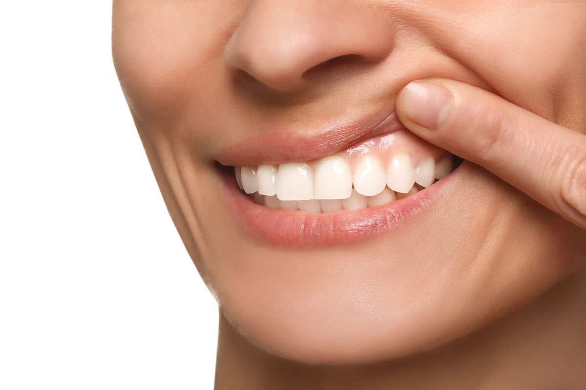
Noticing a white spot, patch, or lump on your gums can be alarming. While many white gum lesions are completely benign and resolve on their own, others can signal underlying conditions that require professional dental evaluation. According to the American Academy of Oral Medicine, white lesions are among the most common oral mucosal abnormalities seen in dental practice, affecting an estimated 5-25% of the adult population at some point in their lives.
In this comprehensive guide, we explain every major cause of white gums -- from common canker sores and oral thrush to more serious conditions like leukoplakia and oral lichen planus. You will learn how to identify each condition, when to seek professional help, what treatments are available, and how much they cost in the US healthcare system. Most importantly, you will understand which symptoms demand immediate attention and which can be safely monitored at home.
What Causes White Spots on Your Gums?
White discoloration of the gums, cheeks, tongue, or palate can arise from dozens of different conditions. The most common causes fall into several major categories:
| Condition | Appearance | Pain Level | Serious? | Self-Resolving? |
|---|---|---|---|---|
| Canker sore (aphthous ulcer) | Small, white-yellow center with red border | Moderate to high | Usually no | Yes (10-14 days) |
| Leukoplakia | Thick, white, hardened patches | None to mild | Can be precancerous | No |
| Oral thrush (candidiasis) | Creamy white patches that can be wiped off | Burning sensation | Moderate (treatable) | No (needs antifungal) |
| Dental cyst/abscess | White-yellow bump near tooth root | Can be severe | Yes (infection) | No |
| Oral lichen planus | Lacy white lines (Wickham striae) | None to moderate | Chronic, needs monitoring | No (manages with treatment) |
| Gingivitis (plaque deposits) | Whitish film along gumline | Mild to moderate | Moderate (can progress) | With improved hygiene |
| Fordyce spots | Tiny white-yellow dots | None | No (normal variant) | Permanent (harmless) |
Let us examine each of these conditions in detail to help you understand what you may be experiencing.
Canker Sores (Aphthous Ulcers)
Canker sores are the most common cause of white lesions in the mouth, affecting approximately 20% of the US population. These small, shallow ulcers appear as round or oval lesions with a white or yellowish center surrounded by a bright red border. They typically develop on the gums, inner cheeks, tongue, or soft palate.
Canker sores are not contagious (unlike cold sores, which are caused by the herpes simplex virus) and are generally harmless, though they can be quite painful -- especially when eating acidic, spicy, or salty foods. Most canker sores heal on their own within 10 to 14 days without scarring.
Common triggers include stress, hormonal changes, minor mouth injuries (from braces or accidental biting), certain foods (citrus, tomatoes, chocolate), nutritional deficiencies (iron, vitamin B12, folate, zinc), and immune system suppression.
Good to Know: Over-the-counter treatments like benzocaine-based gels (Orajel, Anbesol), Debacterol, or prescription mouth rinses containing dexamethasone can significantly reduce canker sore pain and accelerate healing. If you experience canker sores more than three times per year, consider having your dentist test for nutritional deficiencies, which are an underdiagnosed but easily correctable cause.
Leukoplakia: When White Patches Require Monitoring
Leukoplakia presents as thick, white, hardened patches on the gums, inner cheeks, or floor of the mouth. Unlike canker sores, leukoplakia patches cannot be scraped off and are usually painless. The condition is most commonly associated with tobacco use (both smoking and smokeless tobacco) and chronic alcohol consumption.
The critical concern with leukoplakia is its potential for malignant transformation. According to the American Cancer Society, approximately 5-17% of leukoplakia cases undergo dysplastic changes that can progress to oral squamous cell carcinoma over time. The risk is highest for lesions that:
- Are located on the floor of the mouth or the lateral border of the tongue
- Have a non-homogeneous (speckled or verrucous) appearance
- Persist for more than two weeks despite removing the irritant (tobacco, sharp tooth edge)
- Show histological dysplasia on biopsy
"Any white patch in the mouth that persists for more than two weeks without an obvious cause should be evaluated by a dental professional. While the majority of leukoplakia cases are benign, a biopsy is the only way to rule out dysplasia or early-stage oral cancer. Early detection is critical -- the 5-year survival rate for oral cancer detected at Stage I is over 80%, compared to less than 40% for Stage IV."
Warning: If you use tobacco products (cigarettes, cigars, chewing tobacco, snuff) and notice persistent white patches on your gums or anywhere in your mouth, schedule a dental evaluation immediately. Tobacco-related leukoplakia carries the highest risk of malignant transformation. Quitting tobacco dramatically reduces your risk -- the ADA offers resources at ADA.org/tobacco for cessation support.
Oral Thrush (Candidiasis)
Oral thrush is a fungal infection caused by the overgrowth of Candida albicans, a yeast that naturally lives in the mouth in small amounts. When the oral microbiome is disrupted, Candida can proliferate, creating creamy white lesions on the gums, tongue, inner cheeks, and palate. Unlike leukoplakia, thrush lesions can often be wiped away with gauze, revealing red, irritated tissue beneath.
Risk factors for oral thrush include:
- Recent antibiotic use (which kills beneficial bacteria that keep Candida in check)
- Inhaled corticosteroid use (common in asthma patients)
- Diabetes (poorly controlled blood sugar feeds yeast growth)
- Weakened immune system (HIV/AIDS, chemotherapy, organ transplant patients)
- Wearing poorly fitting dentures
- Dry mouth (xerostomia) from medications or medical conditions
- Infancy -- oral thrush is extremely common in newborns and infants
Treatment typically involves antifungal medications such as nystatin oral suspension (swish and swallow), clotrimazole troches, or in severe cases, oral fluconazole. Most cases resolve within 7 to 14 days of treatment.
Dental Cysts and Abscesses
A white or yellowish bump on the gum near a tooth root may indicate a dental cyst or the early stages of a dental abscess. These lesions form when a tooth infection -- typically from an untreated cavity or failed root canal -- creates a pocket of pus or fluid at the root tip.
Initially, a dental cyst may be painless, appearing as a small, firm lump on the gum. However, as the infection progresses, it can evolve into a full-blown abscess with severe throbbing pain, facial swelling, fever, and a foul taste from pus drainage. A dental granuloma may also present as a persistent gum bump.
Treatment requires addressing the underlying tooth infection through root canal therapy, apicoectomy, or extraction. Antibiotics alone cannot cure a dental abscess because the infection source remains inside the tooth.
Warning: A dental abscess is a medical emergency that requires prompt treatment. If you have a white or swollen bump on your gum accompanied by fever, facial swelling, difficulty swallowing, or difficulty breathing, seek emergency dental or medical care immediately. An untreated abscess can spread to the neck, brain, or bloodstream, causing life-threatening complications.
Oral Lichen Planus
Oral lichen planus is a chronic inflammatory condition affecting the oral mucosa. It often presents as a network of fine, lacy white lines (called Wickham striae) on the gums, inner cheeks, or tongue. Some forms also produce red, eroded areas or painful ulcerations.
Oral lichen planus affects approximately 1-2% of the adult population and is most common in women over 40. Its exact cause is unknown, but it is believed to be an autoimmune disorder in which the body's T-cells attack the cells of the oral mucosa. Triggers may include stress, certain medications (NSAIDs, beta-blockers, ACE inhibitors), hepatitis C infection, and allergic reactions to dental materials.
While oral lichen planus is not curable, it can be managed with topical corticosteroids (triamcinolone, fluocinonide), calcineurin inhibitors (tacrolimus, pimecrolimus), and retinoids. Regular monitoring is important because oral lichen planus carries a small (approximately 1-2%) risk of malignant transformation over time.
"Oral lichen planus requires long-term management, not a one-time fix. I recommend that my patients with this condition have oral screenings every 6 months to monitor for any changes in the lesions. The key is to control inflammation, reduce symptoms, and catch any concerning changes early. Most patients do very well with topical steroid therapy and stress management."
Gingivitis and Periodontal Disease
While gingivitis is typically associated with red, swollen gums, it can also cause white or pale-appearing gum tissue. Bacterial plaque buildup along the gumline can create a whitish film, and in severe cases, the gum tissue may appear pale due to reduced blood flow from chronic inflammation.
Additional gum-related causes of white or pale gums include:
- Anemia (iron deficiency) -- Insufficient red blood cells cause pale or whitish-appearing gums throughout the mouth. This is a systemic condition requiring medical evaluation and iron supplementation.
- Chemical burns -- Improper use of teeth whitening products, aspirin placed directly on gums for toothache relief, or accidental exposure to household chemicals can cause white, burned-appearing gum tissue.
- Post-surgical healing -- After tooth extraction, deep scaling, or gum surgery, the healing tissue often appears white or yellowish during the first week of recovery. This is normal and not a cause for concern.
When to See a Dentist: Red Flags You Should Not Ignore
While many white gum lesions are harmless, certain signs should prompt you to schedule a dental appointment promptly:
- Any white patch lasting longer than 2 weeks without improvement
- White lesions that cannot be wiped off (as opposed to food debris or thrush)
- White spots accompanied by pain, bleeding, or numbness
- White lesions with irregular borders or mixed white-and-red areas (erythroleukoplakia)
- A lump or thickening on the gum that changes in size over time
- Difficulty chewing, swallowing, or moving your tongue or jaw
- Unexplained tooth loosening near the white lesion
- Recurrent canker sores (more than 3 episodes per year) may indicate an underlying condition
Good to Know: The Oral Cancer Foundation reports that approximately 54,540 Americans will be diagnosed with oral or oropharyngeal cancer in 2026. The single most effective screening method is a thorough oral examination by a dental professional, which should be part of every routine dental visit. Many dentists now use VELscope or similar fluorescence-based screening devices that can detect precancerous changes invisible to the naked eye.
Diagnosis and Treatment Options
Your dentist will use several diagnostic approaches to determine the cause of white gum lesions:
- Visual and tactile examination -- The dentist examines the size, shape, texture, and location of the lesion and palpates for any hardness or tenderness.
- Medical history review -- Tobacco use, medications, recent illnesses, and immune status provide critical diagnostic clues.
- Culture or swab -- For suspected thrush, a swab is taken and cultured to confirm Candida overgrowth.
- Biopsy -- For persistent or suspicious lesions, a small tissue sample is removed under local anesthesia and sent to a pathology laboratory for microscopic examination. This is the gold standard for ruling out dysplasia or cancer.
- Blood tests -- To check for anemia, nutritional deficiencies, diabetes, or hepatitis C, which can all contribute to oral lesions.
Treatment is condition-specific:
| Condition | Primary Treatment | Typical Cost (Without Insurance) |
|---|---|---|
| Canker sores | OTC gels, prescription rinses, laser treatment | $5 - $30 OTC; $50 - $150 Rx |
| Leukoplakia | Biopsy, tobacco cessation, monitoring, excision if dysplastic | $200 - $600 (biopsy); $500+ (excision) |
| Oral thrush | Antifungal medication (nystatin, fluconazole) | $15 - $80 (prescriptions) |
| Dental abscess | Root canal, extraction, antibiotics | $700 - $2,000+ |
| Oral lichen planus | Topical corticosteroids, monitoring | $30 - $100/month (ongoing) |
| Gingivitis/plaque buildup | Professional scaling, improved home care | $100 - $300 (cleaning) |
Prevention: How to Keep Your Gums Healthy
Many causes of white gum lesions are preventable through consistent oral hygiene and healthy lifestyle choices:
- Brush twice daily with ADA-accepted fluoride toothpaste using a soft-bristled toothbrush. Replace your brush every 3 months.
- Floss daily or use a water flosser to remove plaque and bacteria from between teeth and along the gumline.
- Avoid tobacco in all forms -- cigarettes, cigars, pipes, chewing tobacco, and vaping. Tobacco is the primary risk factor for leukoplakia and oral cancer.
- Limit alcohol consumption -- Heavy drinking increases the risk of oral mucosal damage and leukoplakia.
- Eat a balanced, nutrient-rich diet -- Include foods rich in iron, vitamin B12, folate, and zinc to prevent nutritional deficiencies that trigger canker sores.
- Manage stress -- Chronic stress weakens the immune system and triggers canker sores, lichen planus flare-ups, and thrush.
- See your dentist regularly -- The ADA recommends at least annual dental visits with periodic oral cancer screenings. If you use tobacco or alcohol heavily, consider twice-yearly screenings.
- Practice denture hygiene -- If you wear dentures, remove and clean them daily, and have them checked for proper fit annually to prevent irritation and thrush.
"The mouth is a window to overall health. Changes in gum color, texture, or the appearance of new lesions can be early indicators of not just oral diseases but systemic conditions like anemia, diabetes, or even HIV. I always encourage patients to report any changes they notice in their mouth -- no matter how minor they may seem. A five-minute oral exam can literally save a life."
Frequently Asked Questions About White Gums
What diseases can cause white gums?
The most common conditions causing white gums include canker sores (aphthous ulcers), leukoplakia, oral thrush (candidiasis), oral lichen planus, gingivitis, dental cysts or abscesses, and chemical or thermal burns. Systemic conditions such as iron-deficiency anemia can also cause pale or whitish gums throughout the mouth. In rare cases, white or mixed white-red patches can indicate precancerous dysplasia or early-stage oral cancer, particularly in tobacco and alcohol users.
Are white spots on gums always painful?
No. Many white gum lesions are completely painless, which can create a false sense of security. Leukoplakia, Fordyce spots, early-stage dental cysts, and early oral lichen planus are typically painless. Canker sores, on the other hand, are usually quite painful. The absence of pain does not indicate that a lesion is harmless -- painless white patches that persist for more than two weeks should be evaluated by a dentist to rule out precancerous changes.
Can white gum spots indicate oral cancer?
Yes, though most white gum lesions are benign. Leukoplakia -- persistent white patches that cannot be scraped off -- carries a 5-17% risk of malignant transformation. Erythroleukoplakia (mixed white and red patches) has an even higher risk. The American Cancer Society recommends that any white patch persisting longer than two weeks be evaluated by a dental professional. A biopsy is the definitive method for ruling out dysplasia or cancer. Risk factors include tobacco use, heavy alcohol consumption, HPV infection, chronic UV exposure (lip lesions), and age over 55.
How much does it cost to treat white gum lesions?
Costs vary dramatically depending on the diagnosis. Over-the-counter canker sore treatments cost $5-$30. Prescription antifungal medications for thrush cost $15-$80. A dental office biopsy typically costs $200-$600. Root canal therapy for an abscess ranges from $700-$1,800. Professional scaling and cleaning for gingivitis-related white deposits costs $100-$300. Most dental insurance plans cover diagnostic procedures like biopsies and X-rays at 80-100% after deductible. FSA and HSA funds can be used for all dental treatments.
Can I treat white spots on my gums at home?
Minor canker sores can be managed at home with OTC topical gels (benzocaine, hydrogen peroxide rinses) and will heal on their own within 10-14 days. Rinsing with a warm saltwater solution (1/2 teaspoon salt in 8 oz warm water) can reduce inflammation and promote healing. However, if white spots persist beyond two weeks, are growing, bleed, or are accompanied by pain or other symptoms, you should see a dentist rather than continuing home treatment. Self-diagnosing and delaying professional evaluation for persistent lesions can lead to delayed diagnosis of serious conditions.
Sources
- 1. American Academy of Oral Medicine. "White Lesions of the Oral Mucosa: Clinical Guidelines." AAOM Position Paper, 2024.
- 2. American Cancer Society. "Oral Cavity and Oropharyngeal Cancer: Key Statistics." Cancer.org, 2025.
- 3. Oral Cancer Foundation. "Oral Cancer Facts." OralCancerFoundation.org, 2025.
- 4. American Dental Association. "Oral Health Topics: Canker Sores." ADA.org, 2025.
- 5. Villa A, et al. "Oral Leukoplakia Remains a Challenging Diagnosis." Oral Diseases, 2022;28(1):18-29.
- 6. Centers for Disease Control and Prevention. "Candida Infections of the Mouth, Throat, and Esophagus." CDC.gov, 2024.
- 7. National Institute of Dental and Craniofacial Research. "Oral Lichen Planus." NIDCR.NIH.gov, 2024.
- 8. Fair Health Consumer. "Dental Procedure Cost Estimates." FairHealthConsumer.org, 2025.
- 9. American Association of Oral and Maxillofacial Surgeons. "Oral Biopsy Guidelines." AAOMS Clinical Practice, 2024.